A benchmark for hypothalamus segmentation on T1-weighted MR images
NeuroImage, Volume 264, Issue 1, December 2022, 119741
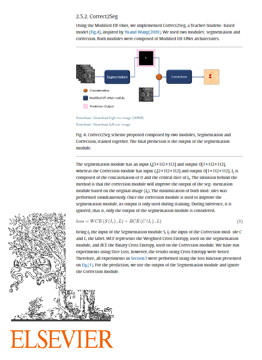
The hypothalamus is a small brain structure that plays essential roles in sleep regulation, body temperature control, and metabolic homeostasis. Hypothalamic structural abnormalities have been reported in neuropsychiatric disorders, such as schizophrenia, amyotrophic lateral sclerosis, and Alzheimer’s disease. Although mag- netic resonance (MR) imaging is the standard examination method for evaluating this region, hypothalamic morphological landmarks are unclear, leading to subjec- tivity and high variability during manual segmentation. Due to these limitations, it is common to find contradicting results in the literature regarding hypothalamic volumetry. To the best of our knowledge, only two automated methods are available in the literature for hypothalamus segmentation, the first of which is our previous method based on U-Net. However, both methods present performance losses when predicting images from different datasets than those used in training. Therefore, this project presents a benchmark consisting of a diverse T1-weighted MR image dataset comprising 1381 subjects from IXI, CC359, OASIS, and MiLI (the latter created specifically for this benchmark). All data were provided using automatically generated hypothalamic masks and a subset containing manually annotated masks. As a baseline, a method for fully automated segmentation of the hypothalamus on T1-weighted MR images with a greater generalization ability is presented. The pro- posed method is a teacher-student-based model with two blocks: segmentation and correction, where the second corrects the imperfections of the first block. After using three datasets for training (MiLI, IXI, and CC359), the prediction performance of the model was measured on two test sets: the first was composed of data from IXI, CC359, and MiLI, achieving a Dice coefficient of 0.83; the second was from OASIS, a dataset not used for training, achieving a Dice coefficient of 0.74. The dataset, the baseline model, and all necessary codes to reproduce the experiments are available at https://github.com/MICLab-Unicamp/HypAST and https://sites.google.com/ view/calgary-campinas-dataset/hypothalamus-benchmarking. In addition, a leaderboard will be maintained with predictions for the test set submitted by anyone working on the same task.
Full paper here: https://doi.org/10.1016/j.neuroimage.2022.119741
Publication Info
- Category: Brain |
- Authors: Livia Rodrigues, Thiago Junqueira Ribeiro Rezende, Guilherme Wertheimer, Yves Santos, Marcondes França, Leticia Rittner
- How to cite: Rodrigues, L.; Rezende, T. J. R.; Wertheimer, G.; Santos, Y.; França, M.; Rittner, L. A benchmark for hypothalamus segmentation on T1-weighted MR images. NeuroImage, v.264, 2022, 119741.
- Published: December 2022


